A photoemission electron microscope (PEEM) produces images using electrons emitted via the photoelectric effect. Since emission depends on light, we can use PEEM to image standing EM waves, such as those produced by diffraction or interference (between light or surface plasmons). The advantage gained is the ability to image photonic phenomena with the high spatial resolution of an electron microscope.
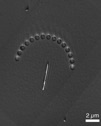
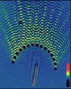
As an example, consider a thin film of conductive indium tin oxide (ITO). In the UV, the film is opaque, but in the visible it is transparent and can guide light. We can see the guided light as interference with the incident laser in PEEM. To do PEEM in the low energy but interesting visible or infrared requires an ultrafast pulse laser (pulse length ~ 100 fs). Short pulses have high photon densities that can overcome the very low likelyhood that two or more photons with sub-work function energy will team up to generate a photoelectron.

UV–PEEM of a semicircle of holes milled by focused ion beam into ITO thing film on glass. Three gold nanowires add a stem to the umbrella.

Two photon–PEEM of the interference of guided blue light scattered into the ITO thin film through the FIB-milled holes.
PSU has a tradition of electron microscope development, which was established by the late Gertrude Rempfer in the 1970’s. Her partnership with O. Hayes Griffiths of UofO led to many refinements to electrostatic optics and a breakthrough in aberration correction for photoemisison electron microscopes.
The key idea is that by combining a lens with an electrostatic mirror, one can simultaneously correct the spherical and chromatic aberrations generated by the accelerating field and objective lens. De-magnification of the image by an intermediate lens brings these aberrations into the right magnitude to be cancelled by the aberrations of the mirror, which are of opposite sign.
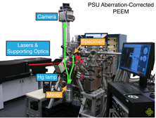
Our work was the realization of the instrument as invisioned by Prof. Rempfer and its subsequent expansion from a tool for life sciences into a tool for the study of surface optical fields. We built the instrument almost entirely on-site and augmented it with CW and ultrafast laser sources. Improvements to instrument continue, including to the core aberration corrector.

PSU aberration-corrected PEEM built entirely in house. The lasers and support optics are on the left. The electron microscope is in the center. High voltage power supplies and computer are on the right and rear.
Unique properties of nanoscale materials make possible interesting takes on more familiar electrical devices such as light emitting diodes, photovoltaics, and antennas. The design process requires a lot of trial and error as nanoscale materials throw surprises at you as a rule.
For example, zinc oxide nanowires can be inexpensively grown on flexible conductive substrate, sealed in insulating cross-linked polystyrene, and coated with conductive polymers to create flexible UV-visible LEDs. Using the same basic design but with added light absorbing CdSe nanoparticles we can instead make a solar cell. Nanoscale metal antennas made with a FIB and relying on plasmonics concentrate light energy to scales well below the wavelength of light — offering exciting possibilities for shrinking optical devices, enhancing existing designs, or creating entirely new technologies.

A solar cell made of zinc oxide nanowires, CdSe particles, conducting polymer, and gold on ITO substrate.

Standing wave electric field of a metal disk antenna excited by off-normal laser light.
The surfaces of cell membranes are incredibly complex, built on a lipid bilayer foundation and packed with proteins that carry out many functions necessary for life. As physicists, we wonder how we can use electrokinesis—that is motion in fluid under an applied electric field—to understand the structure of biomembranes and artificial lipid membranes.
One tool is a class of molecules that are hydrophobic yet carry charge. Many such molecules are well-known drugs and toxins and are interesting in their own right. We can use lipophillic ions to change the surface change of a membrane, which changes the force it feels in an applied field. While the experiments may seem straightforward, understanding changes in electrokinetic fluid flow requires significant efforts in theoretical thinking and computational physics.

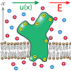
A simple model of a calcium pump imbedded in a lipid membrane and surrounded by electrolyte. In an electric field E, the fluid flows with velocity u(x) that depends on the potential profile of the membrane.

A sarcoplasmic reticulum membrane is packed with with calcium pumps. In the cartoon above the pumps are red disks packed so closely that very little of the membrane (green) is visible.