Crystallographic Image Processing
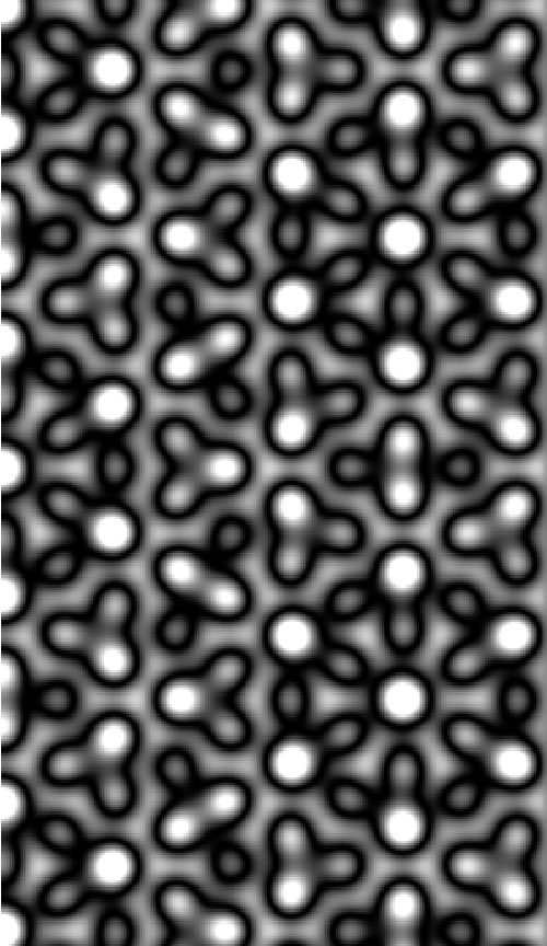
I have been collaborating with PSU Physics faculty member Dr. Peter Moeck, to lay out the theoretical basis for why crystallographic image processing (CIP) techniques allow us to glean information that has been obscured by multiple tips in scanning probe microscopy (SPM). We find that the currents from the two tips differ in a phase term in reciprocal space that can reduce the amplitudes (at a given reciprocal space point) for certain double tip separations, thereby obscuring the current map. CIP lessens this effect by averaging the amplitude and phase at such a point with amplitudes and phases at symmetry-related points.
Structural Fingerprinting of Nanocrystals
In a second collaboration with Dr. Moeck I have derived a formula for the estimation of crystallite thicknesses from precession electron diffraction patterns. This will allow us to develop a procedure for assessing the nanocrystal area that a precessing electron beam illuminates. This, in turn, will be used for structural fingerprinting and electron crystallography of nanocrystals.
Structural Fingerprinting of Nanocrystals
I am also collaborating with PSU Physics faculty member Dr. Erik Sanchez on field enhancement for near-field imaging. My contribution includes a theoretical analysis of tip profiles.
